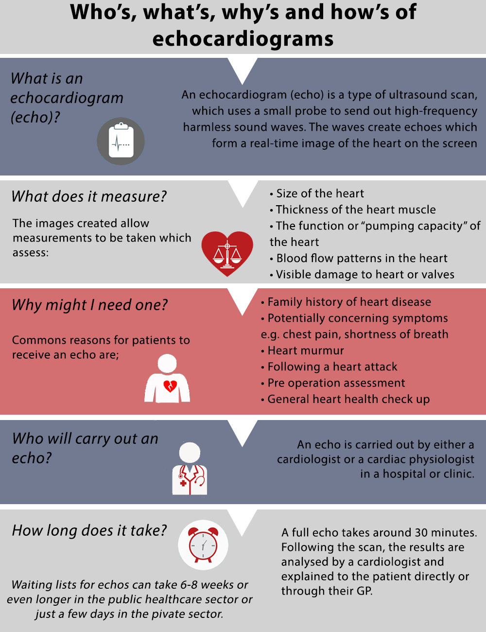
Whether you’ve been referred for a heart scan by your GP or you just want to put your mind at ease about your heart health, an echocardiogram provides a pain-free way to build a complete picture of your heart and quickly evaluate how it’s working.
What is an echocardiogram?
An echocardiogram, or ‘Echo’, is an ultrasound scan. This means that it is very similar to the scans we are familiar with women receiving during pregnancy in order to image and examine a developing baby.
High-frequency, harmless sound waves known as ultrasound waves are directed at the heart through a probe. When the waves reach the heart they bounce back off the structure of the heart, creating ‘echoes’. These echoes are then received again by the probe and processed to form a real-time image of the heart on the screen beside the patient.
What does it measure?
The images created allow measurements to be taken assessing the size of the heart, the function or “pumping capacity” of the heart, thickness of the heart muscle, blood flow patterns in the heart and any visible damage to the heart or the valves.
Why might I need one?
Patients receive echocardiograms for a range of different reasons. These commonly include:
– Family history of heart disease
– Potentially concerning symptoms e.g. chest pain, shortness of breath
– If you have been told you have a heart murmur
– Following a heart attack
– Pre-operation assessment
– General check-up of heart health
Who will carry out an echocardiogram?
An echocardiogram is carried out by either a cardiologist or a cardiac physiologist in a hospital or clinic.
How long does it take?
A full echocardiogram can usually be completed within 30 minutes. Following the scan, the results are analysed by an expert cardiologist and explained to the patient either directly or through a local GP.
Waiting lists for echocardiograms can sometimes be 6–8 weeks or longer in the public healthcare sector or just a few a days in the private sector.
